
Entire pneumorrhachis due to isolated head trauma
To view the full text, please login as a subscribed user or purchase a subscription. Click here to view the full text on ScienceDirect.
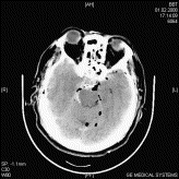
Fig. 1
Computed tomography scan illustrating air and blood images. Also, paranasal sinus bone fractures can be seen. February 1, 2008.
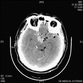
Fig. 2
Computed tomography scan illustrating air and blood images. Also, paranasal sinus bone fractures can be seen. February 1, 2008.
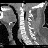
Fig. 3
Computed tomography scan illustrating air at the cervical subarachnoid space.
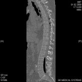
Fig. 4
Computed tomography scan illustrating air at the thoracic and lumbar subarachnoid spaces.
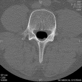
Fig. 5
Computed tomography scan illustrating air in the horizontal plane at level lumbar 4 vertebrae.
To access this article, please choose from the options below
Purchase access to this article
Claim Access
If you are a current subscriber with Society Membership or an Account Number, claim your access now.
Subscribe to this title
Purchase a subscription to gain access to this and all other articles in this journal.
Institutional Access
Visit ScienceDirect to see if you have access via your institution.
