Search Results
200 results
Number of results per page. Upon selecting an option this page will automatically refresh to update the list of articles to your number selected.Fig. 1
Body region injured.
Fig. 1
Body region injured.
Fig. 2
Body region injured by team type, maneuver attempted, and mechanism of injury.
Fig. 2
Body region injured by team type, maneuver attempted, and mechanism of injury.
Fig. 1
Epidemiology of DSP patients.
Fig. 1
Epidemiology of DSP patients.
Fig. 1
A superficial partial-thickness (second-degree) burn. Note presence of blisters with surrounding area of erythema.
Fig. 1
A superficial partial-thickness (second-degree) burn. Note presence of blisters with surrounding area of erythema.
Fig. 2
A deep partial-thickness (second-degree) burn. Note presence of pink-colored dermal appendages within burn.
Fig. 2
A deep partial-thickness (second-degree) burn. Note presence of pink-colored dermal appendages within burn.
Fig. 3
A full-thickness (third-degree) burn.
Fig. 3
A full-thickness (third-degree) burn.
Fig. 1
Anatomy of the trigeminal nerve. Courtesy of Dr Anthony Kaufmann, Winnipeg Centre for Cranial Nerve Disorders, Winnipeg, Manitoba, Canada.
Fig. 1
Anatomy of the trigeminal nerve. Courtesy of Dr Anthony Kaufmann, Winnipeg Centre for Cranial Nerve Disorders, Winnipeg, Manitoba, Canada.
Fig. 2
Red eye in patient with anterior uveitis. Reproduced with permission from the Department of Ophthalmology and Visual Sciences, The Chinese University of Hong Kong, Sept 2002. (For interpretation of the references to color in this figure legend, the reader is referred to the web version of this article.)
Fig. 3
Slit lamp examination in a patient with HZO. Epithelial keratitis may have a dendritic appearance mimicking herpes simplex virus keratitis (A) and stains with fluorescein dye (B). Reproduced with permission from Dr Saad Shaikh.
Fig. 3
Slit lamp examination in a patient with HZO. Epithelial keratitis may have a dendritic appearance mimicking herpes simplex virus keratitis (A) and stains with fluorescein dye (B). Reproduced with permission from Dr Saad Shaikh.
Fig. 4
Slit lamp examination of a patient with stromal keratitis as a result of herpes zoster virus infection. Subepithelial infiltrates are located in the anterior stroma below areas of previous epithelial keratitis. Reproduced with permission from Dr Saad Shaikh.
Fig. 4
Slit lamp examination of a patient with stromal keratitis as a result of herpes zoster virus infection. Subepithelial infiltrates are located in the anterior stroma below areas of previous epithelial keratitis. Reproduced with permission from Dr Saad Shaikh.
Fig. 5
Zoster retinitis characterized by peripheral patches of retinal necrosis. Reproduced with permission from Dr Saad Shaikh.
Fig. 5
Zoster retinitis characterized by peripheral patches of retinal necrosis. Reproduced with permission from Dr Saad Shaikh.
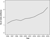
Fig. 1
Total ED visits for abscesses by year 1996 to 2005.
Fig. 1
Total ED visits for abscesses by year 1996 to 2005.
Fig. 2
Distribution of abscess visits by age, 2005.
Fig. 2
Distribution of abscess visits by age, 2005.
Fig. 3
Distribution of abscess visits by body region, 2005.
Fig. 3
Distribution of abscess visits by body region, 2005.
Fig. 1
Injury rates per 100 000 United States resident population by age and sex.
Fig. 1
Injury rates per 100 000 United States resident population by age and sex.
Fig. 2
Body region injured and type of injury sustained during falls from balconies, by sex and age group, United States, 1990-2006.
Fig. 2
Body region injured and type of injury sustained during falls from balconies, by sex and age group, United States, 1990-2006.
Fig. 1
ED visit rate (per 1000 US population) of IDs: 2000-2010.
Fig. 1
ED visit rate (per 1000 US population) of IDs: 2000-2010.
Fig. 2
Trends of antimicrobial prescriptions in EDs for each of the infectious categories of interest.
Fig. 2
Trends of antimicrobial prescriptions in EDs for each of the infectious categories of interest.
Fig. 2
Trends of antimicrobial prescriptions in EDs for each of the infectious categories of interest.
Fig. 2
Trends of antimicrobial prescriptions in EDs for each of the infectious categories of interest.
Fig. 1
Location of injury in postmenopausal (n = 61) and premenopausal victims (n = 1014) with anogenital trauma.
Fig. 1
Location of injury in postmenopausal (n = 61) and premenopausal victims (n = 1014) with anogenital trauma.


