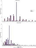
Search Results
750 results
Number of results per page. Upon selecting an option this page will automatically refresh to update the list of articles to your number selected.Fig. 4
Cardiovascular magnetic resonance: the examination was acquired with a 1.5-T machine and dedicated multielement phased-array coil, with cardiac-respiratory trigger/gating and Turbo Spin Echo-T2 Weighted sequences, without and with fat suppression and functional estimate through Steady-State Free Precession multiphase sequences. The examination was completed with the administration of gadolinium via e.v. The Short T1 Inversion Recovery 2-chamber (A), 3-chamber (B), and short-axis (C) sequences show soft edema/inflammation (white arrow) of the left ventricle at the apical segments of the anterior wall, of the septum, and of the apex because of the recent consequence of transient left ventricular apical ballooning syndrome. The Cardiovascular Magnetic Resonance excludes also the presence of any thrombus in the left or right ventricle.
Fig. 4
Cardiovascular magnetic resonance: the examination was acquired with a 1.5-T machine and dedicated multielement phased-array coil, with cardiac-respiratory trigger/gating and Turbo Spin Echo-T2 Weighted sequences, without and with fat suppression and functional estimate through Steady-State Free Precession multiphase sequences. The examination was completed with the administration of gadolinium via e.v. The Short T1 Inversion Recovery 2-chamber (A), 3-chamber (B), and short-axis (C) sequences show soft edema/inflammation (white arrow) of the left ventricle at the apical segments of the anterior wall, of the septum, and of the apex because of the recent consequence of transient left ventricular apical ballooning syndrome. The Cardiovascular Magnetic Resonance excludes also the presence of any thrombus in the left or right ventricle.
Fig. 1
Respiratory rate reporting.
Fig. 1
Respiratory rate reporting.
Figure
Cumulative rates of respiratory depression by fentanyl dose.
Figure
Cumulative rates of respiratory depression by fentanyl dose.
Fig. 1
Flow chart of patients enrolled in the study. RF indicates respiratory failure; CF, cardiovascular failure; Resp, respiratory; Meta, metabolic.
Fig. 1
Flow chart of patients enrolled in the study. RF indicates respiratory failure; CF, cardiovascular failure; Resp, respiratory; Meta, metabolic.
Fig. 4
Inferior vena cava diameter collapses during the respiratory cycle suggesting low CVP.
Fig. 4
Inferior vena cava diameter collapses during the respiratory cycle suggesting low CVP.
Fig. 5
Mortality in respiratory sepsis (left) and other than respiratory sepsis (right).
Fig. 5
Mortality in respiratory sepsis (left) and other than respiratory sepsis (right).
Fig. 1
Early diagnosis of hypothyroidism in old patients presenting with type 2 respiratory failure. ICU indicates intensive care unit.
Fig. 1
Early diagnosis of hypothyroidism in old patients presenting with type 2 respiratory failure. ICU indicates intensive care unit.
Fig. 3
The influence of measured prearrest vital signs, presence of sock or respiratory insufficiency on survival indexed by cause of the arrest in patients resuscitated from asystole or PEA witnessed by the EMS.
Fig. 3
The influence of measured prearrest vital signs, presence of sock or respiratory insufficiency on survival indexed by cause of the arrest in patients resuscitated from asystole or PEA witnessed by the EMS.
Fig. 2
Receiver operating characteristic curve. Respiratory variation of IVC diameter showed good diagnostic accuracy with area under curve of 0.96.
Fig. 2
Receiver operating characteristic curve. Respiratory variation of IVC diameter showed good diagnostic accuracy with area under curve of 0.96.
Fig. 1
Distribution of baseline heart (A) and respiratory rates (B) in the sample of patients with acute asthma (n = 1192).
Fig. 1
Distribution of baseline heart (A) and respiratory rates (B) in the sample of patients with acute asthma (n = 1192).
Fig. 2
The influence of measured prearrest vital signs, presence of shock or respiratory insufficiency on survival indexed by presumed cause of the arrest in patients resuscitated from VF or VT witnessed by the EMS.
Fig. 2
The influence of measured prearrest vital signs, presence of shock or respiratory insufficiency on survival indexed by presumed cause of the arrest in patients resuscitated from VF or VT witnessed by the EMS.
Fig. 1
Emergency department respiratory syndrome visits of patients aged 13 years or older during the month before January 19, 2005 (ED preparedness drill at 3 participating hospitals and citywide).
Fig. 1
Emergency department respiratory syndrome visits of patients aged 13 years or older during the month before January 19, 2005 (ED preparedness drill at 3 participating hospitals and citywide).
Fig. 2
Correlation between the Paco2-Petco2 gradient and respiratory rate. R = 0.21; Y = 1.18 + 0.34X.
Fig. 2
Correlation between the Paco2-Petco2 gradient and respiratory rate. R = 0.21; Y = 1.18 + 0.34X.
Fig. 1
Inferior vena cava diameter changes with respiratory cycle. Inferior vena cava imaged in the same patient without movement of the ultrasound probe. The top image demonstrates maximum AP diameter during expiration with measurement taken as demonstrated by the arrow. Hepatic veins are also seen in this view (arrowhead). The bottom image shows collapse of the IVC (arrow) during inspiration, which has also led to collapse of hepatic veins (arrowhead).
Fig. 2
Forced expiratory volume in the first second (percentage of predicted), heart rate, respiratory rate, and Sao2 values over 3 hours of treatment according to final outcome of patients (■, discharged patients; ●, admitted patients). Brackets represent 1 SD. ⁎Repeated-measures ANOVA.
Fig. 2
Forced expiratory volume in the first second (percentage of predicted), heart rate, respiratory rate, and Sao2 values over 3 hours of treatment according to final outcome of patients (■, discharged patients; ●, admitted patients). Brackets represent 1 SD. ⁎Repeated-measures ANOVA.
Fig. 2
A, Forest plot and heterogeneity of studies comparing ketofol with propofol in “respiratory complications requiring intervention” outcome. B, Funnel plot of studies comparing ketofol with propofol in “respiratory complications requiring intervention” outcome.
Fig. 2
A, Forest plot and heterogeneity of studies comparing ketofol with propofol in “respiratory complications requiring intervention” outcome. B, Funnel plot of studies comparing ketofol with propofol in “respiratory complications requiring intervention” outcome.
Fig. 2
Interleukin 10 cytokine concentrations. Increased levels of IL-10 among both influenza groups (★) compared with bacterial pneumonia (PNA) and other respiratory infections. Stacked bars are means with error bars depicting 95% confidence intervals.
Fig. 2
Interleukin 10 cytokine concentrations. Increased levels of IL-10 among both influenza groups (★) compared with bacterial pneumonia (PNA) and other respiratory infections. Stacked bars are means with error bars depicting 95% confidence intervals.
Figure
Diagram of enrolled for subjects presenting to an inner city ED during the 2012-2013 influenza season with an acute respiratory illness and criteria to indicate antiviral treatment according to CDC guidelines.
Figure
Diagram of enrolled for subjects presenting to an inner city ED during the 2012-2013 influenza season with an acute respiratory illness and criteria to indicate antiviral treatment according to CDC guidelines.
Fig. 1
Relationships between the SLESS and the variables of interest. A, SLESS vs MEDS (r = 0.53; P < .0001). B, SLESS vs SAPS3 (r = 0.55; P < .0001). C, SLESS vs Pao2/Fio2 ratio (r = −0,62; P < .0001). D, SLESS vs respiratory rate (RR) (r = 0.45; P = .0003).
Fig. 1
Relationships between the SLESS and the variables of interest. A, SLESS vs MEDS (r = 0.53; P < .0001). B, SLESS vs SAPS3 (r = 0.55; P < .0001). C, SLESS vs Pao2/Fio2 ratio (r = −0,62; P < .0001). D, SLESS vs respiratory rate (RR) (r = 0.45; P = .0003).
Fig. 1
Association between the number of vital-sign abnormalities and community-acquired pneumonia diagnoses. The histogram plots the percentage of patients diagnosed with CAP as the number of vital-sign abnormalities increases. The number of patients in the sample with each level of abnormality is listed along with the level of abnormality. The following were considered abnormal values for vital signs: temperature higher than 100.4°F; pulse rate higher than 100 beats per minute; respiratory rate greater than 20 breaths per minute; and oxygen saturation less than 95%. The total number of patients is 3252 (1212 patients are not included because of missing data, primarily oxygen saturation).


