
To view the full text, please login as a subscribed user or purchase a subscription. Click here to view the full text on ScienceDirect.
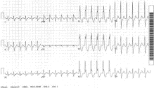
Fig. 1
Regular wide complex tachycardia without distinct P waves and with tall R wave in lead V1.
Fig. 2
Prior ECG study from patient in Fig 1 —normal sinus rhythm and a baseline right bundle branch block (RBBB). Most notably, the Fig 1 ECGs QRS morphology in lead V1 was identical to the current QRS morphology. A diagnosis of supraventricular tachycardia with RBBB was made.
Fig. 3
The typical morphology of the QRS complex in V1 in RBBB is the classic triphasic rsR' pattern. The second peak (R') should be taller than the first peak (r). In contrast, ventricular ectopy—or ventricular tachycardia—arising from the left ventricle generally produces a taller second R in V1.
Fig. 4
The ECG shows normal sinus rhythm with RBBB (rsR'-taller second R wave [the R'] component in V1), but there is also a ventricular ectopic beat (the second QRS complex seen in lead V1) arising from the left ventricle (Rsr'-taller first R wave [the R] component V1).
Fig. 5
Wide complex tachycardia with a rate of 130 and tall R wave in lead V1. Atrioventricular dissociation (best seen in the rhythm strip) is also seen. This ECG is consistent with ventricular tachycardia.
Fig. 6
After therapy with procainamide, the patient's repeat ECG (from Fig 5 ) shows a junctional rhythm with a rate of 54 and AV dissociation. The tall RV1 in lead V1 is not apparent.
Fig. 7
ECG of a 56-year-old woman that was initially misdiagnosed as having SVT with RBBB because of the slightly higher right peak of the QRS complex in V1. The patient developed hemodynamic decompensation after treatment with diltiazem. Closer attention to the ECG reveals the presence of AV dissociation (best seen in leads V1 to V3 before the second, fourth, and fifth QRS complexes), confirming VT.
Fig. 8
An example of left VT with the Rs wave lead V1 morphology.
Fig. 9
ECG from a patient with dehydration and hypokalemia. It shows sinus bradycardia with RBBB and left ventricular bigeminy. The ventricular ectopic beats (ie, with widened QRS complexes) have an Rs morphology in lead V1, in contrast to the sinus-RBBB beats which have the classic triphasic rsR' shape.
Fig. 10
Normal sinus rhythm with evidence of infarction in the inferior and lateral leads. A tall RV1 (R/S ratio > 1) with ST segment depression in V1-V3 was also noted, suggesting posterior wall infarction.
Fig. 11
An ECG from earlier in the year (for comparison with Fig 10 ECG) confirms the diagnosis of an old inferior MI, but the lateral changes and the abnormalities noted in leads V1-V3 were absent.
Fig. 12
This same patient (from Figs 10 and 11 ) had resolution of the ST segment depression at hospital discharge and no further signs of acute ischemia, but persistence of the tall R waves in V1-V3, indicative of the posterior MI. When considered from the posterior perspective, these R waves are actually Q waves.
Fig. 13
Posterior wall myocardial infraction is seen here in anterior leads V1 to V3 with promient R waves, horizontal ST segment depression, and upright T waves. Posterior leads V8 and V9 reveal ST segment elevation with Q waves.
Fig. 14
This ECG is notable for sinus tachycardia, tall R waves in the right precordial leads, right axis deviation, and persistence of the S wave in leads V5-V6. The ECG is consistent with right ventricular hypertrophy.
Fig. 15
Electrocardiographic criteria most often used for RVH include R/S ratio in V1 > 1, R/S ratio in V5 or V6 < 1, RV1 > 7 mm, right axis deviation beyond +110 degrees, or a qR pattern in V1. In the presence of a RBBB (as seen in this example) or incomplete RBBB, RVH can be diagnosed by the presence of an R or R' > 10 mm.
Fig. 16
Severe RVH may be associated with sustained delayed repolarization of the right ventricular myocardium, resulting in ST segment depression and inverted T waves in the precordial leads. This is often referred to as right ventricular “strain.”
Fig. 17
This ECG reveals sinus tachycardia with Q waves in the inferior leads, an inverted T wave in lead III, a large S wave in lead I, tall R wave in lead V1, and persistence of the S wave in V5 and V6 (R/S ratio < 1). The patient was diagnosed with pulmonary embolism.
Fig. 18
Another ECG from a patient with PE. Inversion of T waves is not uncommon in the inferior and precordial leads.
Fig. 19
The ECG shows a tall R wave in lead V1 and Q waves in the inferior leads, suggestive of an inferoposterior MI. However, the ECG also shows slight prolongation of the QRS complex, a short PR segment, and slurring of the upstroke of the QRS complex (the delta wave), diagnostic of Wolff-Parkinson-White syndrome.
Fig. 20
Hypertrophic cardiomyopathy (HCM) is often associated with tall RV1. Large depolarization forces, directed in a rightward, anterior, and superior direction, can create QRS complexes in the right precordial leads that are of large amplitude. The resulting QRS complex may have an RS or Rs (see Fig 21 ) morphology. In addition to the large amplitude QRS complexes, another key feature that results from the septal depolarization is the presence of deep but narrow Q waves in the lateral leads (I, AVL, V5-V6) and occasionally in the inferior leads. These Q waves, when combined with tall RV1, may lead to the erroneous diagnosis of inferior and/or lateral MI with posterior extension.
Fig. 21
Hypertrophic cardiomyopathy is often associated with tall R wave in lead V1; depicted here is an example of Rs morphology of the right precordial QRS complexes.
Fig. 22
An example of mirror-image dextrocardia.
Fig. 23
The ECG in Fig 23 was taken in the same patient after the precordial and limb leads were reversed (compared with the ECG in Fig 22 ).
Fig. 24
This ECG was mistakenly interpreted as showing evidence of a previous posterior MI because of the tall R wave in lead V1. The “backwards” R wave progression in leads V1-V3 and the isolated T wave inversion in lead V3 suggest that leads V1 and V3 have been reversed. Comparison with a previous ECG (see Fig 25 ) confirms this suspicion.
Fig. 25
Confirmation of lead reversal in the ECG from Fig 24 .
Fig. 26
This ECG was mistakenly interpreted as showing evidence of a previous anterior MI because of the poor R wave progression in lead V3. However, this “backwards” R wave progression and the isolated T wave flattening in lead V3 once again suggest that leads V1 and V3 have been reversed. An even stronger clue is the presence of a biphasic P wave in lead V3. Inverted or biphasic P waves are very common in lead V1, but are rare in the other precordial leads in the absence of an ectopic rhythm.
Abstract
Tall lead V1 (tall RV1), defined as an R/S ratio equal to or greater than 1, is not an infrequent occurrence in emergency department patients. This electrocardiographic finding exists as a normal variant in only 1% of patients. Physicians should therefore be familiar with the differential diagnosis for this important QRS configuration. The electrocardiographic entities which can present with this finding include right bundle branch block, left ventricular ectopy, right ventricular hypertrophy, acute right ventricular dilation (acute right heart strain), type a Wolff-Parkinson-White syndrome, posterior myocardial infarction, hypertrophic cardiomyopathy, progressive muscular dystrophy, dextrocardia, misplaced precordial leads, and normal variant. Various cases are presented to highlight the different causes of the tall RV1. (Am J Emerg Med 2001;19: 504-513. Copyright © 2001 by W.B. Saunders Company)
To access this article, please choose from the options below
Purchase access to this article
Claim Access
If you are a current subscriber with Society Membership or an Account Number, claim your access now.
Subscribe to this title
Purchase a subscription to gain access to this and all other articles in this journal.
Institutional Access
Visit ScienceDirect to see if you have access via your institution.
