
Sixty-four–slice computed tomographic coronary angiography: will the “triple rule out” change chest pain evaluation in the ED?

To view the full text, please login as a subscribed user or purchase a subscription. Click here to view the full text on ScienceDirect.
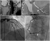
Fig. 1
The 64-slice CTCA of a patient with risk factors for coronary disease who presented with atypical chest pain. Multiplanar reformat images demonstrate a moderate stenosis in the left anterior descending artery (A, arrow) and moderate disease in the circumflex artery (B, arrow). The right coronary artery has moderate plaque throughout the proximal and midportions (C, arrowheads) and a critical lesion in the crux of the distal right coronary artery (D, arrow). These lesions are demonstrated on invasive cardiac catheterization (E, F).
Fig. 2
The 64-slice CTCA of a patient who presented with chest pain and a mildly abnormal stress test. Three-dimensional imaging of the heart (A) and coronary artery tree (B) demonstrates a left dominant coronary system. Multiplanar reformat images of the left anterior descending (C), left main and proximal diagonal (D), circumflex (E), and right coronary (F) arteries demonstrate no plaque of any kind. The patient was reassured, and no further cardiac evaluation was necessary.
Fig. 3
Multislice CT of the heart, aorta, and proximal iliac arteries (A), and a close-up view of the heart and proximal aorta (B). This patient presented to the ED with a diagnosis of chest and back pain and was diagnosed with a type B aortic dissection (arrow) by CT angiography.
Fig. 4
Oblique view of a type B aortic dissection. The spiral “flap” is clearly seen (arrows) starting at the takeoff of the left subclavian and dissecting distally through the descending aorta.
Fig. 5
A 51-year-old man presented with chest pain and shortness of breath. Gated CT of the chest detected thrombi of the pulmonary arteries in a classic “saddle embolus” conformation. Axial images (A) and oblique views (B) demonstrate the thrombi (arrows).
Abstract
Sixty-four–slice computed tomographic (CT) coronary angiography is a new technique for the noninvasive visualization of the coronary arteries. It enables noninvasive detection of coronary plaque and determination of severity without instrumentation of the heart. Although not yet commonly used in the emergency department setting, it stands poised to dramatically change the way that patients with chest pain are evaluated.
In addition to evaluation of the coronary arteries, CT angiography has long been used to evaluate patients for other dangerous causes of chest pain such as aortic dissection and pulmonary embolus. Although these new scanners excel at all of these diagnostic modalities, the true excitement is in the possibility of combining several different protocols into one, allowing for multiple causes of chest pain to be “ruled out” simultaneously.
This article describes the current state of the art of cardiac CT, current state of research, and current areas of controversy.
To access this article, please choose from the options below
Purchase access to this article
Claim Access
If you are a current subscriber with Society Membership or an Account Number, claim your access now.
Subscribe to this title
Purchase a subscription to gain access to this and all other articles in this journal.
Institutional Access
Visit ScienceDirect to see if you have access via your institution.
