
Emergency physicians can easily obtain ultrasound images of anatomical landmarks relevant to lumbar puncture☆

To view the full text, please login as a subscribed user or purchase a subscription. Click here to view the full text on ScienceDirect.
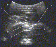
Fig. 1
Paramedian approach image obtained with curved array probe (a, lamina; b, ligamentum flavum; c, epidural space; d, dura mater; e, subarachnoid space).
Fig. 2
Paramedian approach image obtained with a linear array probe (a, laminae; b, ligamentum flavum; c, dura mater; d, subarachnoid space; e, epidural space).
Fig. 3
Midline approach image obtained with linear array probe (a, spinous processes; b, dura mater/ligamentum flavum; c, subarachnoid space).
Fig. 4
Vertebral body illustration, midline and paramedian approaches.
Fig. 5
Spinal anatomy illustration, midline and paramedian approaches.
Fig. 6
Physician perception of ease of landmark palpation.
Abstract
Introduction
Although ultrasound has been used in administering epidural anesthesia, it is unknown if emergency physicians (EPs) can obtain ultrasound images useful for lumbar puncture.
Objective
The objective of the study was to determine EPs' ability to apply a standardized ultrasound technique for visualizing landmarks surrounding the dural space.
Methods
Two EPs sought to identify relevant anatomy in emergency patients. Visualization time for 5 anatomical structures (spinous processes or laminae, ligamentum flavum, dura mater, epidural space, subarachnoid space), body mass index, and perception of landmark palpation difficulty were recorded.
Results
Seventy-six subjects were enrolled. Soft tissue and bony anatomical structures were identified in all subjects. Mean body mass index was 31.4 ± 9.8 (95% confidence interval, 29.1-33.6). High-quality images were obtained in less than 1 minute in 153 (87.9%) scans and in less than 5 minutes in 174 (100%) scans. Mean acquisition time was 57.19 seconds; SD, 68.14 seconds; range, 10 to 300 seconds.
Conclusion
In this cohort, EPs were able to rapidly obtain high-quality ultrasound images relevant to lumbar puncture.
To access this article, please choose from the options below
Purchase access to this article
Claim Access
If you are a current subscriber with Society Membership or an Account Number, claim your access now.
Subscribe to this title
Purchase a subscription to gain access to this and all other articles in this journal.
Institutional Access
Visit ScienceDirect to see if you have access via your institution.
This project was presented at the American College of Emergency Physicians Research Forum, October 2004.
☆This project did not receive support in the form of equipment, drugs, or grants.
