
Spontaneous hemorrhagic angiomyolipoma present with massive hematuria leading to urgent nephrectomy
To view the full text, please login as a subscribed user or purchase a subscription. Click here to view the full text on ScienceDirect.
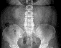
Fig. 1
The x-ray image of the abdomen region. The image shows the abnormal kidney on the left with dark patches, which are a hematoma.
Fig. 2
Abdominal sonography. The picture shows a hyperechoic mass in the left renal region.
Fig. 3
Computed tomography showed a 3 × 5-cm, fat-containing tumor noted at the left lower pole of the kidney, with hematoma in the left perirenal space.
Fig. 4
The tumor was composed of a mixture of proliferating vessels, fat cells, and spindle smooth muscle cell–like cells.
Abstract
Abstract
Managing acute abdomen is a challenge for physicians in the emergency department. An immediate surgery is sometimes required for the hemorrhagic uropathy of renal angiomyolipoma. We present a case with left flank pain and findings of hematuria and pyuria. Abdominal and pelvic computed tomographic scans showed a 3 × 5-cm, fat-containing tumor at the left lower pole of the kidney and a bleeding and perirenal hematoma in the left perirenal space. An urgent left nephrectomy was lifesaving for the postoperative finding of renal angiomyolipoma. It is important to consider the possibility of the presence of such a tumor when spontaneous retroperitoneal hemorrhage and hypovolemic shock are encountered.
To access this article, please choose from the options below
Purchase access to this article
Claim Access
If you are a current subscriber with Society Membership or an Account Number, claim your access now.
Subscribe to this title
Purchase a subscription to gain access to this and all other articles in this journal.
Institutional Access
Visit ScienceDirect to see if you have access via your institution.
