
Validation of a porcine comb burn model

To view the full text, please login as a subscribed user or purchase a subscription. Click here to view the full text on ScienceDirect.
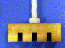
Fig. 1
The brass comb used to induce thermal contact burns.
Fig. 2
Appearance of burn wound created by brass comb immediately after injury.
Fig. 3
Appearance of burn wound created by brass comb 1 day after injury.
Fig. 4
Appearance of burn wound created by brass comb 1 week after injury.
Fig. 5
Photomicrograph of interspace 7 days after injury demonstrating necrosis in ischemic unburned interspace. Hematoxylin-eosin stain.
Abstract
Objective
A brass comb burn model that creates 3 full-thickness burns separated by 3 interspaces of unburned skin representing the zone of ischemia has been described in rats. We evaluated this model in pigs.
Methods
Design—observational. Subjects—6 pigs (20-25 kg). Interventions—4 burns created on each animal on the dorsum using a brass comb with 4 rectangular prongs preheated in boiling water and applied for 30 seconds resulting in 4 rectangular 10 × 20–mm full-thickness burns separated by three 5 × 20–mm unburned interspaces. Outcomes—wounds observed at 1, 2, 3, and 7 days for evidence of necrosis in unburned interspaces. Full-thickness biopsies from interspaces were evaluated with hematoxylin-eosin staining 7 days after injury for evidence of necrosis. Data analysis—the percentages of interspaces progressing to necrosis were analyzed with descriptive statistics.
Results
Twenty-four comb burns with 72 unburned interspaces were created evenly distributed between the animals. The percentages of interspaces that progressed to full-thickness necrosis at 1, 2, 3, and 7 days after injury were 88.9% (64/72; 95% confidence interval [CI], 79.6%-94.3%), 88.9% (64/72; 95% CI, 79.6%-94.3%), 88.9% (64/72; 95% CI, 79.6%-94.3%), and 97.7% (70/72; 95% CI, 90.4%-99.2%), respectively. There was perfect agreement between gross inspection and histomorphology.
Conclusions
The comb burn model in swine results in the progression of most unburned ischemic interspaces to full-thickness necrosis within 1 to 7 days.
To access this article, please choose from the options below
Purchase access to this article
Claim Access
If you are a current subscriber with Society Membership or an Account Number, claim your access now.
Subscribe to this title
Purchase a subscription to gain access to this and all other articles in this journal.
Institutional Access
Visit ScienceDirect to see if you have access via your institution.
