
Reappraisal of radiographic signs of pneumoperitoneum at emergency department

To view the full text, please login as a subscribed user or purchase a subscription. Click here to view the full text on ScienceDirect.
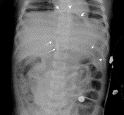
Fig. 1
A 1-year-old female subject with cecal perforation due to chronic inflammation. The KUB shows Rigler sign (broken arrows), falciform ligament sign (straight arrows), and cupola sign (arrowheads).
Fig. 2
A 28-year-old female subject with perforated duodenal ulcer. The KUB shows triangle sign (broken arrows) and fissure for ligament teres sign (straight arrows).
Fig. 3
An 83-year-old male subject with perforated duodenal ulcer. The CXR shows hyperlucent liver sign (broken arrows) and dolphin sign (straight arrows).
Fig. 4
A 68-year-old male subject with proximal jejunum perforation due to ischemic bowel disease. The CXR shows anterior superior oval sign (broken arrows) and left anterior superior oval sign (straight arrows).
Fig. 5
A 76-year-old male subject with pylorus perforation. The KUB shows doge cap sign (arrows).
Fig. 6
A 77-year-old male subject with Billroth II anastomosis perforation. The KUB shows hepatic edge sign (broken arrows) and football sign (straight arrows).
Fig. 7
A 73-year-old male subject with perforated gastric ulcer. The KUB shows inverted V sign (broken arrows) and focal radiolucency (straight arrows).
Fig. 8
A 72-year-old male subject with perforated peptic ulcer. The KUB shows urachus sign (arrows).
Fig. 9
A 72-year-old female subject with perforated gastric ulcer. The CXR shows bilateral subphrenic radiolucency (arrows).
Abstract
Purpose
This study aimed to evaluate the sensitivities of the reported free air signs on supine chest and abdominal radiographs of hollow organ perforation. We also verified the value of supine radiographic images as compared with erect chest and decubitus abdominal radiographs in detection of pneumoperitoneum.
Methods
Two hundred fifty cases with surgically proven hollow organ perforation were included. Five hundred twenty-seven radiographs were retrospectively reviewed on the picture archiving and communication system. Medical charts were reviewed for operative findings of upper gastrointestinal tract, small bowel, or colon perforations. The variable free air signs on both supine abdominal radiographs (KUB) and supine chest radiographs (CXR) were evaluated and determined by consensus without knowledge of initial radiographic reports or final diagnosis. Erect CXR and left decubitus abdominal radiographs were evaluated for subphrenic free air or air over nondependent part of the right abdomen.
Result
Upper gastrointestinal tract perforation was proven in 91.2%; small bowel perforation, in 6.8%; and colon perforation, in 2.0%. The positive rate of free air was 80.4% on supine KUB, 78.7% on supine CXR, 85.1% on erect CXR, and 98.0% on left decubitus abdominal radiograph. Anterior superior oval sign was the most common radiographic sign on supine KUB (44.0%) and supine CXR (34.0%). Other free air signs ranged from 0% to 30.4%.
Conclusions
Most free air signs on supine radiographs are located over the right upper abdomen. Familiarity with free air signs on supine radiographs is very important to emergency physicians and radiologists for detection of hollow organ perforation.
To access this article, please choose from the options below
Purchase access to this article
Claim Access
If you are a current subscriber with Society Membership or an Account Number, claim your access now.
Subscribe to this title
Purchase a subscription to gain access to this and all other articles in this journal.
Institutional Access
Visit ScienceDirect to see if you have access via your institution.
