
Correlation of sonographic measurements of the internal jugular vein with central venous pressure

To view the full text, please login as a subscribed user or purchase a subscription. Click here to view the full text on ScienceDirect.
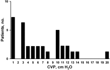
Fig. 1
Distribution of patients according to CVP, which ranged from 1 to 20 cm H2O.
Fig. 2
Comparison of CVP and AP diameter with patient in supine position and at the end of expiration.
Abstract
Determination of volume status is crucial in treating acutely ill patients. This study examined bedside ultrasonography of the internal jugular vein (IJV) to predict central venous pressure (CVP). Ultrasonography was performed on 34 nonventilated patients with monitored CVPs. The IJV was measured during the respiratory cycle and with the patient in different positions. Mean IJV diameter in patients with CVP less than 10 cm H2O was 7.0 mm (95% confidence interval [CI], 5.7-8.3) vs 12.5 mm (95% CI, 11.2-13.8) in patients with CVP of 10 cm H2O and greater. Measurement of end expiratory diameter with the patient supine had the highest correlation coefficient: 0.82 (95% CI). There was strong agreement among ultrasonographers: correlation coefficient, 0.92 (95% CI). This pilot study shows promise that ultrasonography of the IJV can be a noninvasive tool to predict CVP. Measurement of end expiratory diameter in supine patients exhibited a high correlation to CVP.
To access this article, please choose from the options below
Purchase access to this article
Claim Access
If you are a current subscriber with Society Membership or an Account Number, claim your access now.
Subscribe to this title
Purchase a subscription to gain access to this and all other articles in this journal.
Institutional Access
Visit ScienceDirect to see if you have access via your institution.
