Body surface mapping vs 12-lead electrocardiography to detect ST-elevation myocardial infarction
Affiliations
- Internal Medicine Virginia Commonwealth University Health System, PO Box 980401, Richmond, VA 23298-0401, USA
Correspondence
- Corresponding author. Tel.: +1 804 828 5250; fax: +1 804 828 8597.

Affiliations
- Internal Medicine Virginia Commonwealth University Health System, PO Box 980401, Richmond, VA 23298-0401, USA
Correspondence
- Corresponding author. Tel.: +1 804 828 5250; fax: +1 804 828 8597.
Affiliations
- Craigavon Cardiac Centre, Craigavon, Northern Ireland BT63 5XD, UK
Affiliations
- Internal Medicine Virginia Commonwealth University Health System, PO Box 980401, Richmond, VA 23298-0401, USA
Affiliations
- Internal Medicine Virginia Commonwealth University Health System, PO Box 980401, Richmond, VA 23298-0401, USA
Affiliations
- Regional Medical Cardiology Centre, Royal Victoria Hospital Belfast, Northern Ireland BT12 6BA, UK
Affiliations
- University of Vermont College of Medicine, Burlington, VT 05401, USA
- Maine Medical Center, Portland, ME 04102, USA
Affiliations
- Regional Medical Cardiology Centre, Royal Victoria Hospital Belfast, Northern Ireland BT12 6BA, UK
To view the full text, please login as a subscribed user or purchase a subscription. Click here to view the full text on ScienceDirect.

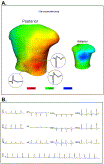
Fig. 1
Eighty-lead BSM ECG (A) and 12-lead ECG (B) from a study subject. The 80-lead BSM ECG map shows an area of ST (J point) elevation (maximum = +0.68 mm) over the lower right posterior chest wall and an area of ST depression (minimum = −1.49 mm) over the left anterolateral chest wall, in keeping with acute posterior MI. Underlying ECG traces from the posterior electrodes with ST elevation are displayed. The accompanying 12-lead ECG shows no significant ST elevation and only minor nonspecific ST depression in leads V3, V4, and V5.
Abstract
A prospective, multicenter trial was conducted in patients with nontraumatic chest pain in 4 hospitals to determine whether an 80-lead body surface map electrocardiogram system (80-lead BSM ECG) improves detection of ST-segment elevation in acute myocardial infarction (STEMI) compared with a standard 12-lead electrocardiogram (ECG) in an emergency department (ED) setting. A trained ED or cardiology staff member (technician or nurse) recorded a 12-lead ECG and 80-lead BSM ECG from each subject at initial presentation. Serial biomarkers (total creatine kinase [CK], CK-MB, and/or troponin) were obtained according to individual hospital practice. Of the 647 patients evaluated, 589 had available biomarkers results. Eighty-lead BSM ECG improved detection of biomarker-confirmed STEMI compared with the 12-lead ECG for CK-MB–defined STEMI (100% vs 72.7%, P = .031; n = 364) or troponin-defined STEMI (92.9% vs 60.7%, P = .022; n = 225). Specificity for STEMI was high (range, 94.9%-97.1%) with no significant difference between 80-lead BSM ECG and 12-lead ECG. Right ventricular involvement complicating inferior STEMI was detected by 80-lead BSM ECG in 2 (22%) of 9 patients with CK-MB–defined MI and in 2 (22%) of 9 patients with troponin-defined MI. The infarct location missed most commonly on 12-lead ECG but detected by 80-lead BSM ECG was inferoposterior MI. We conclude that BSM using 80-lead BSM ECG is more sensitive for detection of STEMI than 12-lead ECG, while retaining similar specificity.
To access this article, please choose from the options below
Purchase access to this article
Claim Access
If you are a current subscriber with Society Membership or an Account Number, claim your access now.
Subscribe to this title
Purchase a subscription to gain access to this and all other articles in this journal.
Institutional Access
Visit ScienceDirect to see if you have access via your institution.
Related Articles
Searching for related articles..


