
Preexcitation syndromes: diagnostic consideration in the ED

To view the full text, please login as a subscribed user or purchase a subscription. Click here to view the full text on ScienceDirect.
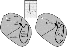
Fig. 1
The impulse formation and conduction process in a patient with normal sinus rhythm. “A” denotes transmission of the impulse through intraatrial pathways to the AV node. “B” denotes transmission of the impulse through the AV node and into the His-Purkinje system. “C” represents the propagation of the impulse through the ventricular myocardium via the specialized conduction system. The ECG complex is the surface electrical manifestation of this process with a normal PR interval and QRS complex, exhibiting the normal conduction through the atria to the AV node (“A” and “B”) and ventricles (“C”), respectively.
Fig. 2
The impulse formation and conduction process in a patient with WPW syndrome in normal sinus rhythm. “A” denotes transmission of the impulse through intraatrial pathways to the AV node. “B” denotes transmission of the impulse through the AV node and into the His-Purkinje system. “C” describes the rapid conduction of the impulse through atrial tissues to the accessory pathway, arriving at the ventricle sooner than anticipated had normal transmission occurred via the AV node; this action is manifested as a shortened PR interval on the ECG. “D,” the delta wave, is the segment of ventricular myocardium that depolarizes earlier than anticipated had normal conduction occurred. “E” is the summation or fusion of the electrical waveforms transmitted to the ventricular myocardium via both the accessory pathway and AV node—as depicted by a widened QRS complex.
Fig. 3
Left lateral accessory AV pathway. Subtle delta waves are best seen in leads V3 and V4. A positive delta wave in V1 and the inferior leads (II, III, aVF) along with a negative to isoelectric QRS in lead aVL point to this location in this ECG. This patient was admitted for atrial fibrillation with a rapid ventricular response and wide QRS morphology.
Fig. 4
Right posteroseptal accessory AV pathway. Occasional narrow QRS complexes without evidence of preexcitation are seen (arrows). The QRS and delta waves in the inferior leads are negative during preexcitation, simulating prior inferior myocardial infarction. The right ventricular origin is indicated by the negative delta wave in V1, which promptly becomes upright in V2. Delta waves are best seen in the lateral precordial leads.
Fig. 5
Left posteroseptal accessory AV pathway. As in Fig. 4 , negative delta and QRS deflections are seen in leads III and aVF, mimicking prior inferior myocardial infarction. The left ventricular origin of this pathway is indicated by the positive delta wave in V1.
Fig. 6
The same patient in Fig. 4 , now with suppressed accessory pathway conduction. Procainamide was administered to this patient after an episode of atrial fibrillation with rapid ventricular conduction and a wide QRS complex. Note the inverted T waves in the inferior leads, which are the result of ventricular memory of preexcitation in these leads.
Fig. 7
Wide QRS complex tachycardia (WCT) with retrograde AV node conduction in the patient with WPW syndrome, an ART. This form of WCT uses the accessory pathway as the anterograde limb of the reentry loop (“A”) with the AV node functioning as the retrograde limb (“C”). The reentry loop involves the ventricular (“B”) and atrial (“D”) myocardium. The impulse arrives at the ventricle via the accessory pathway and is propagated distally via the ventricular myocardium; the QRS complex is widened in that the His-Purkinje system is not used to rapidly and efficiently propagate the impulse, thereby producing a wide QRS complex.
Fig. 8
Narrow QRS complex tachycardia (NCT) with anterograde AV node conduction in the patient with WPW syndrome, an orthodromic reciprocating tachycardia (ORT). This form of NCT uses the AV node as the anterograde limb of the reentry loop (“A”) with the accessory pathway functioning as the retrograde limb (“C”). The reentry loop involves the ventricular (“B”) and atrial (“D”) myocardium. The impulse arrives at the ventricle via the AV node and is propagated distally using the His-Perkinje system, thereby producing a narrow QRS complex.
Fig. 9
Atrioventricular reentry tachycardia. A reentry circuit in which an electrical impulse is conducted down the AV node-His-Purkinje axis, forming a narrow QRS complex, and reentering the atria via retrograde conduction through an AV accessory pathway distant from the AV node. Note the distinct p waves following the QRS complex in lead V1 (red arrow; pseudo-r′ waves) and in leads II, III, and AVF (blue arrow; pseudo-S waves), thus creating a “short r-p” tachycardia pattern. This patient manifested ventricular preexcitation on resting ECGs. (For interpretation of the references to color in this figure legend, the reader is referred to the web version of this article.)
Fig. 10
Irregular, wide QRS complex tachycardia consistent with preexcited atrial fibrillation in the patient with WPW syndrome. This form of atrial fibrillation uses the AV node (“A”) and accessory pathway (“B”) for anterograde conduction to the ventricular myocardium. “C” is the segment of ventricular myocardium which depolarizes earlier than anticipated had normal conduction occurred. “D” is the summation or fusion of the electrical waveforms transmitted to the ventricular myocardium via both the accessory pathway and AV node—as depicted by a widened QRS complex. The resultant ECG waveform is an irregular, wide QRS complex tachycardia with delta wave and beat-to-beat variation in QRS complex morphology.
Fig. 11
Preexcited atrial fibrillation in the patient with WPW syndrome. The ECG waveform is an irregular, wide QRS complex tachycardia with delta wave and beat-to-beat variation in QRS complex morphology. The delta wave, not present in all beats, signifies the depolarization of the ventricular myocardium due to the impulse arriving via the accessory pathway. The beat-to-beat variation in the QRS complex results from the varying contribution to ventricular depolarization arriving via the accessory pathway and the AV node.
Fig. 12
Atrial fibrillation with WPW syndrome. Note the variety of QRS morphologies, but the relatively constant amplitude of voltages, in contrast to the wide variation in QRS voltages seen with PVT. A probable fusion beat is seen (arrow).
Fig. 13
Series of ECGs from a patient with WPW syndrome and atrial fibrillation, treated with procainamide (courtesy of Ralph Verdino, MD). A, WPW syndrome with atrial fibrillation on presentation to the ED. Irregular wide QRS complex tachycardia with varying QRS configurations representing varying degrees of accessory pathway mediated ventricular preexcitation and concomitant AV node mediated conduction. This is differentiated from PVT by its relatively nonchaotic pattern and from SVT with aberrant conduction by both the heterogeneity of QRS complexes and the lack of a stereotypical bundle-branch pattern. B, ECG recorded after initiation of treatment with procainamide in the ED. Note intermittent narrow QRS complexes due to Na+ channel–mediated blockade of accessory pathway conduction. C, ECG obtained later that day showing normal sinus rhythm with a narrow QRS complex. The procainamide infusion was discontinued at this time. D, ECG from the following day demonstrating a return of ventricular preexcitation. The patient was subsequently taken to the electrophysiology laboratory for a successful radiofrequency ablation.
Abstract
Preexcitation syndromes are a common cause of paroxysmal tachycardias presenting to the ED. Emergency physicians should be familiar with the common electrocardiographic manifestations of preexcitation, particularly the Wolff-Parkinson-White abnormality, as these conditions require specific therapeutic management. This article reviews the pathophysiology of preexcitation, along with the electrocardiographic findings of Wolff-Parkinson-White and its associated tachyarrhythmias.
To access this article, please choose from the options below
Purchase access to this article
Claim Access
If you are a current subscriber with Society Membership or an Account Number, claim your access now.
Subscribe to this title
Purchase a subscription to gain access to this and all other articles in this journal.
Institutional Access
Visit ScienceDirect to see if you have access via your institution.
