
A comparison of 2 types of chest compressions in a porcine model of cardiac arrest☆

To view the full text, please login as a subscribed user or purchase a subscription. Click here to view the full text on ScienceDirect.
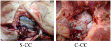
Fig. 1
Methylene blue staining extent of brain tissue after CPR.
Fig. 2
Photographs of histopathologic findings. A, A few neuron cells lost the processus and swelled mildly. B, Most neuron cells lost the processus, swelled heavily, nuclear pycnosis and Nissl bodies lost. A part of the cell membrane is dissolved and neuron cellular necrosis (thin arrow) with the reaction of the neuronophage (thick arrow). C, Few cytoplasms of myocardial cells are deeply stained with nuclear pycnosis (thin arrow). D, Most cytoplasms of myocardial cells are deeply stained with nuclear pycnosis (thin arrow); myocardial interstitial substance are widen and interstitial infiltration of few inflammatory cells (thick arrow). E, Glomeruluses are normal. Renal tubular epithelial cells swelled mildly. F, Glomeruluses are basic normal. Renal tubular epithelial cells swelled, and interstitial infiltration of lots of inflammatory cells, most of which are leukomonocytes (arrow). G, Hepatocytes swelled, sinus hepaticus is broaden with blood congested. H, Hepatocytes swelled, sinus hepaticus is broaden obviously with blood congested(arrow). S-CC: A, C, E, and G; C-CC: B, D, F, and H (H&E ×400).
Abstract
Objective
Chest compressions performed by some medical workers are of poor quality, which are too few and shallow with incomplete release. This study was designed to compare the effects of these clinical quality chest compressions with standard manual chest compressions in a porcine model of cardiac arrest.
Methods
Ventricular fibrillation was induced in 18 pigs by programed electrical stimulation. Then, 40 mg methylene blue was injected into right atrium after 4 minutes of untreated ventricular fibrillation (VF), followed by cardiopulmonary resuscitation for 9 minutes. Defibrillation was attempted at 13 minutes of cardiac arrest. Animals of no restoration of spontaneous circulation after 4 times of defibrillations were announced dead and dissected immediately to observe the cerebral perfusion with methylene blue coloration. Resuscitated animals were executed to remove the tissues of pallium, cardiac muscle, kidney, and liver for histopathology after evaluating a porcine Cerebral Performance Category score at 24 hours after cardiac arrest. All animals were randomized to the following 2 groups: (1) standard manual chest compressions group (n = 9)—chest compression rates were kept at 100 ± 5 cpm and compression depth at 50 ± 1 mm with complete release by Heartstart MRx Monitor; (2) clinical quality chest compressions group (n = 9)—chest compression rates were kept at 80 ± 5 cpm and compression depth at 37 ± 1 mm with incomplete release.
Results
Compared with clinical quality chest compressions, standard manual chest compressions produced greater restoration of spontaneous circulation, neurologically normal 24-hour survival, and histopathologic findings.
Conclusions
High-quality chest compressions improve outcomes of resuscitation, especially postresuscitation brain damage.
To access this article, please choose from the options below
Purchase access to this article
Claim Access
If you are a current subscriber with Society Membership or an Account Number, claim your access now.
Subscribe to this title
Purchase a subscription to gain access to this and all other articles in this journal.
Institutional Access
Visit ScienceDirect to see if you have access via your institution.
☆The study was supported by capital medical science development scientific research fund (NO 2005-1006). We also appreciate professor Ai-Hu Wang who provided technical help in inducing VF with programed electrical stimulation instrument.
