
Left ventricular “temporary pacemaker wire”: a viable option in emergency situation
To view the full text, please login as a subscribed user or purchase a subscription. Click here to view the full text on ScienceDirect.
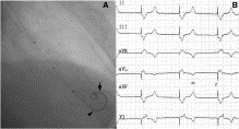
Fig. 1
A, Anteroposterior cardiac fluoroscopic view showing the BMW wire with nontraumatic J-tipped configuration (arrow) advanced into the LV apex. Note the 3 radiopaque markers of the Spectranetics Quick-Cross catheter in the LV and its tip near the apex (arrowhead). This catheter advanced up to the tip of the wire will provide support and insulation through the entire length of the BMW guidewire in the body. B, Postpacing electrocardiogram: 6-lead rhythm strip showing the pacing electrocardiogram. Note the QRS complex in the V1, leads 2, 3, and avF, suggesting LV pacing.
To access this article, please choose from the options below
Purchase access to this article
Claim Access
If you are a current subscriber with Society Membership or an Account Number, claim your access now.
Subscribe to this title
Purchase a subscription to gain access to this and all other articles in this journal.
Institutional Access
Visit ScienceDirect to see if you have access via your institution.
