
Ultrasound diagnosis of papilledema and increased intracranial pressure in pseudotumor cerebri

To view the full text, please login as a subscribed user or purchase a subscription. Click here to view the full text on ScienceDirect.
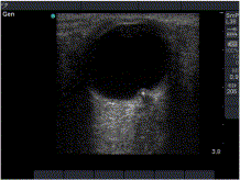
Fig. 1
A rounded hyperechoic mass (asterisk) arising from the optic disc and protruding into the posterior chamber, consistent with papilledema.
Fig. 2
Optic nerve sheath diameter of 7.1 mm, consistent with increased intracranial pressure. Note that the measurement is performed 3.0 mm posterior to the globe.
Abstract
Bedside ultrasound has been used to identify intracranial hypertension through the measurement of optic nerve sheath diameter. This case report describes the sonographic detection of papilledema and a wide optic nerve sheath in a patient with pseudotumor cerebri who presented to the Emergency Department with headache and photophobia, and in whom fundoscopy was poorly tolerated. Bedside ultrasound may represent an alternate means of assessing for papilledema when a traditional fundoscopic exam is non-diagnostic.
To access this article, please choose from the options below
Purchase access to this article
Claim Access
If you are a current subscriber with Society Membership or an Account Number, claim your access now.
Subscribe to this title
Purchase a subscription to gain access to this and all other articles in this journal.
Institutional Access
Visit ScienceDirect to see if you have access via your institution.
