T-wave inversion: cardiac memory or myocardial ischemia?
VA Ann Arbor/University of Michigan Healthcare System, Ann Arbor, MI, USA
Tiziano M. Scarabelli, MD, PhD
Wayne State University School of Medicine, Detroit, MI, USA
To view the full text, please login as a subscribed user or purchase a subscription. Click here to view the full text on ScienceDirect.

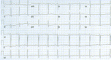
Fig. 1
Baseline normal sinus rhythm.
Fig. 2
Baseline paroxysmal atrial fibrillation.
Fig. 3
Paced rhythm (right ventricular pacing, with occasional AV pacing).
Fig. 4
Postpacing electrocardiogram with TwIs.
Fig. 5
Attenuation of TwI, 2 weeks after pacemaker clinic.
This article presents a case report of a 74-year-old man with T-wave inversion (TwI) in atrial fibrillation noted during routine pacemaker interrogation.
To access this article, please choose from the options below
Purchase access to this article
Claim Access
If you are a current subscriber with Society Membership or an Account Number, claim your access now.
Subscribe to this title
Purchase a subscription to gain access to this and all other articles in this journal.
Institutional Access
Visit ScienceDirect to see if you have access via your institution.
Published by Elsevier Inc.
Access this article on
Visit ScienceDirect to see if you have access via your institution.
Related Articles
Searching for related articles..


