
Acute myocardial infarction with cardiogenic shock in a patient with acute aortic dissection

To view the full text, please login as a subscribed user or purchase a subscription. Click here to view the full text on ScienceDirect.
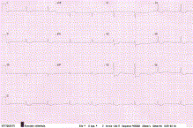
Fig. 1
Electrocardiography showing signs of diffuse myocardial ischemia.
Fig. 2
A, Coronary angiography of the left coronary artery before PCI. Right anterior oblique with cranial angulation. B, Coronary angiography of the left coronary artery after PCI and stenting of the LMCA. Left anterior oblique with cranial angulation.
Fig. 3
A, Transesophageal echocardiography with visualization of an intimal flap. B, Transesophageal echocardiography with visualization of an intimal flap with an eccentric aortic regurgitation.
To access this article, please choose from the options below
Purchase access to this article
Claim Access
If you are a current subscriber with Society Membership or an Account Number, claim your access now.
Subscribe to this title
Purchase a subscription to gain access to this and all other articles in this journal.
Institutional Access
Visit ScienceDirect to see if you have access via your institution.
