Ligamentum flavum hematoma due to stretching exercise: a case report and review of literature
Correspondence
- Corresponding author at: No. 2, Zhongzheng 1st Rd Lingya District, Kaohsiung City 802, Taiwan, ROC. Tel.: +886 774 906 33; fax: +886 774 052 31

Correspondence
- Corresponding author at: No. 2, Zhongzheng 1st Rd Lingya District, Kaohsiung City 802, Taiwan, ROC. Tel.: +886 774 906 33; fax: +886 774 052 31
 Article Info
To view the full text, please login as a subscribed user or purchase a subscription. Click here to view the full text on ScienceDirect.

Fig. 1
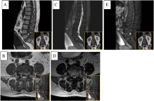
Sagittal and axial T1-weighted (A and B), T2-weighted (C and D) and gadolinium-enhanced T2-weighted (E) magnetic resonance images showing an intrathecal cyst-like lesion with adjacent retrospinal soft tissue enhancement at L4-L5 level with severe thecal sac compression and severe encroachment of the bilateral neural foramina.
Fig. 2
Intraoperative findings showed ligamentum flavum (A, arrow). After dissecting ligamentum flavum, dark liquefied material (B, arrow) was found, and a capsule lesion was identified (C, arrow). Decompression and exposure of dura (D, arrow) was performed.
Fig. 3
Sagittal (A) and axial (B) T2-weighted images of the same area 5 months later showing complete resolution after the patient underwent L4 through L5 laminectomies and removal of intraspinal lesion, which was proven to be a old hemorrhage in fibrous tissue.
To access this article, please choose from the options below
Purchase access to this article
Claim Access
If you are a current subscriber with Society Membership or an Account Number, claim your access now.
Subscribe to this title
Purchase a subscription to gain access to this and all other articles in this journal.
Institutional Access
Visit ScienceDirect to see if you have access via your institution.
Related Articles
Searching for related articles..


