
Cerebral air embolism after central venous catheterization
To view the full text, please login as a subscribed user or purchase a subscription. Click here to view the full text on ScienceDirect.
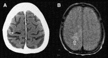
Fig. 1
(A) Brain CT with air bubbles, mainly on the right parietal lobe (thin arrow), indicating cerebral air embolism lodging in the periphery of the middle cerebral arteries. (B) MRI (proton-intensity image) showing cerebral infarction on the right motor cortex (thick arrow).
—Insertion of a central venous (CV) catheter is associated with various complications, although it is a routine procedure in the emergency department and intensive care unit. We report a case of cerebral air embolism during CV catheterization, a rare but potentially fatal complication. Emergency physicians should be familiar with the knowledge on its pathophysiology, presenting symptoms, diagnosis of choice, and treatment.
To access this article, please choose from the options below
Purchase access to this article
Claim Access
If you are a current subscriber with Society Membership or an Account Number, claim your access now.
Subscribe to this title
Purchase a subscription to gain access to this and all other articles in this journal.
Institutional Access
Visit ScienceDirect to see if you have access via your institution.
© 2001 W.B. Saunders Company. Published by Elsevier Inc. All rights reserved.
