
A case of a gas embolism in the aorta induced by necrotizing gastroenteritis
,,,

Department of Traumatology and Critical Care Medicine (CCM), National Defense Medical College (NDMC)
To view the full text, please login as a subscribed user or purchase a subscription. Click here to view the full text on ScienceDirect.
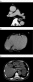
Fig. 1
Trunk CT on arrival. The trunk CT demonstrated a gas embolism in the ascending aorta (A), coronary artery (B), portal vein, and wall of the stomach (C) (white arrow).
A 69-year-old man with epigastralgia and vomiting continuing for 7 hours was transported to this department. At the time of examination, he was drowsy and in a state of shock. He showed a markedly distended abdomen and epigastric tenderness. Arterial blood gas showed hypoxia and severe metabolic acidosis. Computed tomography revealed intramural gas in the wall of the stomach and small intestine, and multiple gas in the portal vein, mesenteric vein, pulmonary artery, coronary artery, and ascending aorta.
To access this article, please choose from the options below
Purchase access to this article
Claim Access
If you are a current subscriber with Society Membership or an Account Number, claim your access now.
Subscribe to this title
Purchase a subscription to gain access to this and all other articles in this journal.
Institutional Access
Visit ScienceDirect to see if you have access via your institution.
© 2008 Elsevier Inc. Published by Elsevier Inc. All rights reserved.
