Calcaneal avulsion fractures: complications from delayed treatment
To view the full text, please login as a subscribed user or purchase a subscription. Click here to view the full text on ScienceDirect.

Fig. 1
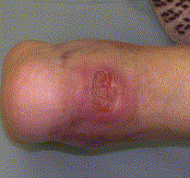
Case 1: open wound on posterior heel developed when the fracture seen in Fig. 2 was not treated expediently. The displaced fragment causes pressure on the overlying skin, leading to necrosis.
Fig. 2
Case 1: radiograph showing the displaced beak fracture. This fracture pattern must be differentiated from more common patterns as a fracture that must be reduced emergently.
Fig. 3
Case 2: a larger fracture fragment in a calcaneal avulsion fracture.
Fig. 4
Skin necrosis seen in case 2.
Fig. 5
Skin necrosis seen in case 3, which was not fixed until 2 days after the injury. At this time, it was too late, and the skin necrosed after the incision. This illustrates the importance of reducing the fragment as soon as it is recognized.
To access this article, please choose from the options below
Purchase access to this article
Claim Access
If you are a current subscriber with Society Membership or an Account Number, claim your access now.
Subscribe to this title
Purchase a subscription to gain access to this and all other articles in this journal.
Institutional Access
Visit ScienceDirect to see if you have access via your institution.
Related Articles
Searching for related articles..


