Endovascular treatment of a bleeding secondary aortoenteric fistula in a high-risk patient☆
To view the full text, please login as a subscribed user or purchase a subscription. Click here to view the full text on ScienceDirect.

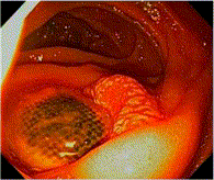
Fig. 1
Preoperative esophagogastroduodenoscopy showing AEF in the duodenum with no evidence of bleeding.
Fig. 2
Coronal computed tomographic angiography demonstrating an intact tubular surgical graft, without perigraft air, contrast extravasation, pseudoaneurysm, or fluid collection. It depicts the length of the right aortoiliac tube graft and the distance from the left renal artery to the AEF. Ascites is present.
Fig. 3
A, Axial image depicts the close contact between the graft and the duodenum, which has no posterior wall. B, Coronal image demonstrating the same.
Fig. 4
A, Postdeployment angiogram confirming exclusion of the fistula. B, Aortouniiliac graft.
Abstract
We report a patient with life-threatening gastrointestinal bleeding caused by a secondary aortoenteric fistula (AEF). Because the patient had severe medical comorbidities, an endovascular approach was chosen for hemorrhage control. Endovascular treatment of aortoenteric fistula provides another treatment option that may be particularly valuable in patients whose comorbidities would preclude open repair.
To access this article, please choose from the options below
Purchase access to this article
Claim Access
If you are a current subscriber with Society Membership or an Account Number, claim your access now.
Subscribe to this title
Purchase a subscription to gain access to this and all other articles in this journal.
Institutional Access
Visit ScienceDirect to see if you have access via your institution.
☆The authors have no financial or other conflicts of interest related to the submission.
Related Articles
Searching for related articles..


