Ischemic-appearing electrocardiographic changes predict myocardial injury in patients with intracerebral hemorrhage☆☆☆★
Affiliations
- Department of Emergency Medicine, Massachusetts General Hospital, Boston, MA 02114, USA
Affiliations
- Department of Emergency Medicine, the University of Utah Hospital, Salt Lake City, UT, USA
Affiliations
- Department of Neurology, Massachusetts General Hospital, Boston, MA 02114, USA
Affiliations
- Department of Neurology, Massachusetts General Hospital, Boston, MA 02114, USA
Affiliations
- Department of Neurology, Massachusetts General Hospital, Boston, MA 02114, USA
Affiliations
- The Calgary Stroke Program, Department of Clinical Neurosciences, University of Calgary, Calgary, Alberta, Canada
Affiliations
- Department of Neurology, Massachusetts General Hospital, Boston, MA 02114, USA
Affiliations
- Department of Neurology, Massachusetts General Hospital, Boston, MA 02114, USA
- The Center for Human Genetic Research, Massachusetts General Hospital, Boston, MA 02114, USA
Affiliations
- Department of Emergency Medicine, Massachusetts General Hospital, Boston, MA 02114, USA
Affiliations
- Department of Emergency Medicine, Massachusetts General Hospital, Boston, MA 02114, USA
Correspondence
- Corresponding author. Tel.: +1 617 7265273; fax: +1 617 7260311.

Affiliations
- Department of Emergency Medicine, Massachusetts General Hospital, Boston, MA 02114, USA
Correspondence
- Corresponding author. Tel.: +1 617 7265273; fax: +1 617 7260311.
 Article Info
To view the full text, please login as a subscribed user or purchase a subscription. Click here to view the full text on ScienceDirect.

Fig. 1
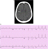
A, Head computed tomographic scan of patient OF on admission. An illustrative patient, OF is a 70-year-old woman with no history of coronary artery disease who presented with confusion. Her head computed tomographic scan showed right temporoparietal ICH. Her ECG showed T wave inversions in leads I, aVL, and V1 to V6. Her peak troponin was 1.8 ng/mL. The patient subsequently underwent a stress test performed with technetium Tc 99m sestamibi that disclosed a modest degree of reversible inferoapical defect. B, Electrocardiogram of patient OF on admission.
Abstract
Objectives
Myocardial injury is common among patients with intracerebral hemorrhage (ICH). However, it is challenging for emergency physicians to recognize acute myocardial injury in this population, as electrocardiographic (ECG) abnormalities are common in this setting. Our objective is to examine whether ischemic-appearing ECG changes predict subsequent myocardial injury in the context of ICH.
Methods
Consecutive patients with primary ICH presenting to a single academic center were prospectively enrolled. Electrocardiograms were retrospectively reviewed by 3 independent readers. Anatomical areas of ischemia were defined as I and aVL; II, III, and aVF; V1 to V4; and V5 and V6. Medical record review identified myocardial injury, defined as troponin I or T elevation (cutoff 1.5 and 0.1 ng/mL, respectively), within 30 days.
Results
Between 1998 and 2004, 218 patients presented directly to our emergency department and did not have a do-not-resuscitate/do-not-intubate order; arrival ECGs and troponin levels were available for 206 patients. Ischemic-appearing changes were noted in 41% of patients, and myocardial injury was noted in 12% of patients. Ischemic-appearing changes were more common in patients with subsequent injury (64% vs 37%; P = .02). After multivariable analysis controlling for age and cardiac risk factors, ischemic-appearing ECG changes independently predicted myocardial injury (odds ratio, 3.2; 95% confidence interval, 1.3-8.2). In an exploratory analysis, ischemic-appearing ECG changes in leads I and aVL as well as V5 and V6 were more specific for myocardial injury (P = .002 and P = .03, respectively).
Conclusion
In conclusion, although a range of ECG abnormalities can occur after ICH, the finding of ischemic-appearing changes in an anatomical distribution can help predict which patients are having true myocardial injury.
To access this article, please choose from the options below
Purchase access to this article
Claim Access
If you are a current subscriber with Society Membership or an Account Number, claim your access now.
Subscribe to this title
Purchase a subscription to gain access to this and all other articles in this journal.
Institutional Access
Visit ScienceDirect to see if you have access via your institution.
☆Presentation information: Abstract presented at SAEM Annual Meeting, Chicago, IL, May 2007.
☆☆Conflicts of Interest Disclosure: Dr Joshua N. Goldstein has received consulting fees from CSL Behring.
★This study is funded by the National Institute of Neurological Disorders and Stroke ( NIH K23NS059774 ).
Related Articles
Searching for related articles..


