
Abdominal oxygen saturation for monitoring return of spontaneous circulation in out-of-hospital cardiac arrest using near infrared spectrophometry
a b s t r a c t
Aim: We used near-infrared spectrophotometry to assess the initial and final abdominal and cerebral saturations during cardiopulmonary resuscitation (CPR) of patients with out-of-hospital cardiac arrest to determine if there is a correlation between increase in these saturation values and return of spontaneous circulation.
Materials and methods: We evaluated 34 patients with out-of-hospital cardiac arrest without witnesses brought to our emergency department. Abdominal and cerebral saturations were measured using near-infrared spectrophotometry from the start of CPR. Cardiopulmonary resuscitation was performed for a maximum of 30 minutes. The effect of abdominal saturations in patients with or without spontaneous circulation restored through CPR was then assessed.
Results: Thirty-four patients (17 males + females) with a mean age of 63.06 +- 11.66 years were included in the study. A significant correlation was determined between increase in abdominal saturations measured at the start and end of CPR and the return of spontaneous circulation (P b .001). A good positive correlation was also identified between abdominal saturation and return of spontaneous circulation.
Conclusion: Patients with increased abdominal and cerebral saturation values have a higher survival rate after appropriate CPR. This Noninvasive measurement system and monitoring of patients during CPR may be a good method of predicting return of spontaneous circulation and assessing abdominal perfusion.
(C) 2014
Despite the development of successful Resuscitation techniques, it is difficult to predict the return of spontaneous circulation (ROSC). Several studies have recently been performed on the practicability of near- infrared spectrophotometry (NIRS) for assessing oxygenation of vital organs such as the brain after cardiac arrest [1-8]. The majority of these have suggested that cardiopulmonary resuscitation (CPR) affects elevated cerebral saturation during resuscitation and can be used to predict ROSC. Oxygenation of other organs is as important as that of the brain during CPR. Near-infrared spectrophotometry can assess myocardi- al, hepatic, and intestinal saturation by measuring tissue saturation [9-11]. Although Cardiac activity can be evaluated with echocardiography, a noninvasive technique, invasive methods such as catheterization need to be used to assess abdominal oxygenation. Near-infrared spectropho- tometry measures total oxygen saturation in a specific tissue volume by
* Corresponding author at: Department of Emergency Medicine, Faculty of Medicine, Recep Tayyip Erdogan University, 53020 Rize, Turkey. Tel.: +90 464 217 0366; fax: +90
464 217 0367.
E-mail address: [email protected] (A. Kalkan).
the approximate assessment of oxygen saturation of the hemoglobin fraction in the terminal vasculature of the target [12]. Continuous moni- toring of abdominal saturation with this noninvasive technique can elicit important therapeutic information. Measures to protect the abdominal organs against hypoxia can thus be taken during and after resuscitation.
Near-infrared spectrophotometry (cerebral oximetry)
Cerebral oximetry is a neurologic and tissue saturation monitoring technique developed for adult and pediatric cardiac surgery in the 1970s. This technology is still used today in areas such as noncardiac surgery, cardiology, resuscitation, trauma, pediatrics, and neurology [13]. Several studies have used it in the prediction of the development of necrotizing enterocolitis in newborns. The INVOS-5100c device with 4 probes can measure cerebral and Tissue oxygenation. Near- infrared spectrophotometry monitors generally include a light source at one side of the optode and one or more detectors at varying distances on the opposite side of the light source. The optode is applied over the region of interest. The detected light curves from the light source to the detector [14]. When it passes through the skin, skull, and tissue,
http://dx.doi.org/10.1016/j.ajem.2014.11.029
0735-6757/(C) 2014
the emitted light is partly scattered, reflected, and absorbed by chromo- phores, such as myoglobin, hemoglobin, cytochromes, water, and mela- nin. The remaining reflected light is collected in the detectors. The difference in absorption of oxygenated and deoxygenated hemoglobin is measured and represents the oxygenation status of the tissue. By ap- plying the modified Beer-Lambert law, numerical values for the mea- surements are displayed on the monitor. This primarily venous oxygen saturation level is a function of local tissue oxygen consumption and, therefore, oxygen delivery, making this measurement a reliable re- flection of perfusion [1].
Cerebral O2 saturation (ScO2) values in normal healthy individuals range from 55% to 75%. If ScO2 values decrease by more than 25% from basal values during cardiovascular surgery under NIRS monitoring, cere- bral ischemia should be suspected and appropriate measures taken [15]. The purpose of this study was to assess by NIRS the initial and final ab- dominal and cerebral saturations during CPR of patients with out-of- hospital CA to determine if there is a correlation between increase in
these saturation values and ROSC.
- Materials and methods
- Study design and setting
In this observational study, data on abdominal tissue oxygen satura- tion were collected from patients experiencing out-of-hospital CA. The abdominal and cerebral saturation values were accessible to the Attending emergency physicians but were not used in any treatment protocol or therapeutic decision. This study was performed at the Emergency Medi- cine Department, Medical Faculty, Recep Tayyip Erdogan University, Turkey, between July and September 2014. The local ethical committee approved the study protocol before commencement. Written consent forms were received from the relatives of all patients included in the study. Our hospital emergency department (ED) admits approximately 120000 patients annually. Approximately 150 to 200 of these patients with CA arrive by ambulance. Because the urban settlement area in our region is small (~250 km2), all cases of CA reach our ED in a relatively short time (10-15 minutes), with the provision of Basic Life Support (BLS) / Advanced life support as required in compliance with Europan Rescucitation Council (ERC) Guidelines 2010.
Participants
Forty-three patients (mean age, 63.06 +- 11.66; range, 21-82 years) experiencing out-of-hospital CA brought by ambulance and lacking witnesses were included in the study. The CA study participants who were older than 18 years had no spontaneous circulation on arrival to the hospital despite receiving CPR at the scene outside the hospital and during transport in the ambulance. Patients with head and abdominal traumas suspected of cerebrovascular pathology and a known history of aortic aneurism were not included in the study. Nine patients were excluded due to the possibility of aortic aneurysm rupture because this was known to be in their medical histories. The duration of the CPR at the scene and during transport had been less than 15 minutes before arrival to the ED, but that of the CA was unknown. The emergency medical services team included a physician and a paramedic. These provided CPR in accordance with ERC Guidelines 2010 on site and during transport as required before admission to the ED. No mechanical compression device was used. Only patients without pulse, despite having received CPR before admission to the ED, were included in this study. Admitted patients without spontaneous circulation continued to receive CPR from the emergency staff. Length of CA before resuscitation was not determined. Monitoring with cerebral and tissue oximetry was provided as soon as the CPR team started resuscitation. Cardiopulmonary resuscitation in line with the American Heart Association (AHA) Ad- vanced Cardiac Life Support (ACLS) 2010 guideline was administered to all patients. Duration of CPR was for a maximum of 30 minutes.
Cerebral and abdominal tissue saturations were monitored until patients were declared dead or until spontaneous circulation returned. The ab- dominal saturation states were evaluated according to ROSC or otherwise. Those with successful ROSC were referred to the intensive care unit.
Cerebral oximetry measurements
Advanced life support was provided for each patient with arrest. Pa- tients were received by a 6-member resuscitation team in the ED. The team consisted of 2 physicians, 2 nurses, and 2 paramedics. One nursing task was to monitor and record cerebral and abdominal tissue saturation. Recording was performed with an INVOS 5100C cerebral/somatic oxime- ter (Covidien, USA). Of the 4 probes of the device, 2 were stuck to the fron- tal region of the head in a banana form. The other 2 were stuck on the abdominal region, with one just over the top-left of the navel and the other to the front abdominal wall immediately above the liver. Recording took place throughout resuscitation. Near-infrared spectrophotometry was not applied before admission because of possible disconnection after unsuccessful CPR interventions.
Statistical analysis
Descriptive statistics are presented as frequency, percentage, mean, SD and median, and minimum and maximum values. Fisher exact test or the Pearson ?2 test was used in the analysis of relations between categorical variables. The Mann-Whitney U test was used in the analysis of differences between the 2 groups’ measurement values. Receiver operating characteristic analysis was performed in the calculation of sensitivity, specificity, and Area under curve values of specific variables in discriminating between surviving and nonsurviving patients. Odds ratios with a 95% confidence interval were set in compar- ing risk of death in groups based on determined cutoff points. P b .05 was regarded as significant. Analyses were performed using SPSS
18.00 (SPSS, Chicago, IL).
- Results
Thirty-four patients with out-of-hospital CA and a mean age of 63.06 +-
11.66 years were included in the study. Both sexes were equal in numbers. Cardiopulmonary resuscitation in line with American Heart Association (AHA) resuscitation norms was administered to all patients. Spontaneous circulation was restored in 13 patients (38.2%), but not in the other 21 (61.8%). Patient characteristics are shown in Table 1. When brought to our ED, patients had pulseless electrical activity (n = 6, 17.6%), ventricular fibrillation (n = 18, 52.9%), and asystolic rhythms (n = 10, 29.4%).
Increases in abdominal O2 saturation (rSO2) and ScO2 values with restoration of spontaneous circulation during CPR were compared in all patients (Table 2). Patients’ cerebral saturations were measured from the right frontal and left frontal regions, and abdominal satura- tions, from the liver and the navel. The highest ScO2 values in the right frontal region of patients with Restored spontaneous circulation were
70.69 +- 11.90, and the lowest ScO2 values were 21.92 +- 8.61. Increases in ScO2 values in the right and left frontal regions between the start of CPR and ROSC were 48.77 +- 14.82 (P b .001) and 47.54 +- 14.26
(P b .001), respectively. The highest and lowest ScO2 values in the right frontal region in patients in whom spontaneous circulation could
Table 1
Patient characteristics
Characteristic n (%)
Age (mean +- SD) 64.09 +- 13.66
Male 17/34 (50)
Initial rhythm (asystole) 10/34 (29.44)
Initial rhythm (VF) 18/34 (52.94)
Initial rhythm (PEA) 6/34 (17.61)
ROSC 13/34 (38.2)
Cerebral (ScO2)/abdominal (rSO2) saturation states of study participants
Area O2 saturation measured ROSC P
Yes (mean +- SD) No (mean +- SD)
|
Lowest right ScO2 |
21.92 (8.61) |
17.90 (4.65) |
.052 |
|
Highest right ScO2 |
70.69 (11.90) |
23.76 (8.63) |
b.001 |
|
Mean right ?ScO2 |
48. 77 (14.82) |
5.86 (6.03) |
b.001 |
|
Lowest left ScO2 |
23.15 (9.96) |
19.19 (6.15) |
.077 |
|
Highest left ScO2 |
70.69 (9.94) |
24.71 (9.41) |
b.001 |
|
Mean left ?ScO2 |
47.54 (14.26) |
5.52 (6.22) |
b.001 |
|
Lowest right rSO2 |
25.15 (8.03) |
21.57 (6.07) |
.248 |
|
Highest right rSO2 |
66.23 (7.30) |
21.29 (6.43) |
b.001 |
|
Mean right ?rSO2 |
41.08 (9.69) |
-0.29 (2.49) |
b.001 |
|
Lowest left rSO2 |
23.15 (6.27) |
21.62 (8.14) |
.431 |
|
Highest left rSO2 |
65.77 (5.61) |
22.57 (8.50) |
b.001 |
|
Mean left ?rSO2 |
42.62 (7.84) |
0.95 (2.25) |
b.001 |
not be restored were 23.76 +- 8.63 and 17.90 +- 4.65, respectively. The levels of increase in ScO2 values in the right and left frontal regions after CPR were 5.86 +- 6.03 and 5.52 +- 6.22, respectively, in which case CPR was terminated and death declared. The highest right and left frontal lobe ScO2 values and the increase in both frontal lobe ScO2 values were significantly higher in the patients in whom spontaneous circulation was restored compared to those without restored circulation (P b .001). No significant difference was determined between the lowest right and left lobe ScO2 values (P N .05) (Table 2). Spontaneous circula- tion was established in patients with an elevation in ScO2 values, but not in patients lacking elevation.
The highest and lowest rSO2 values in the right abdominal region in patients with restored spontaneous circulation were 66.23 +- 7.30 and
25.15 +- 8.03, respectively. Increases in rSO2 values in the right and left abdominal regions in patients with restored spontaneous circula- tion were 41.08 +- 9.69 (P b .001) and 42.62 +- 7.84 (P b .001). The
highest and lowest rSO2 values in the right abdominal region in patients without restored spontaneous circulation were 21.29 +- 6.43 and
21.57 +- 6.07, respectively. Increases in rSO2 values in the right and left abdominal regions when CPR was stopped were – 0.29 +- 2.49 and 0.95 +- 2.25, respectively. The highest right and left rSO2 values and the increase in rSO2 values in both abdominal regions were significantly higher in patients with restored spontaneous circulation compared to those without (P b .001). No significant difference was determined between the lowest right and left rSO2 values (P N .05) (Table 2). Spontaneous circulation was restored in patients with a rise in rSO2 values but not in patients lacking increase.
No significant differences were determined between patients with restored spontaneous circulation and those without in terms of blood glucose levels and blood gas values measured at the start and end of CPR. Receiver operating characteristic analysis was performed in the differentiation of patients with or without restored spontaneous circulation based on the parameters shown in Table 3. Although the duration of CPR exceeded 22 minutes in all nonsurviving patients (sensitivity, 100%), it was less than or equal to 22 minutes in surviving patients (sensitivity, 95.2%). Cutoff values of 35% and 37% were deter- mined for rises in ScO2 levels in the right and left frontal regions, respec- tively. The risk of mortality in patients with a less than or equal to 35% rise in ScO2 in the right frontal region was 14.08 times greater than that in patients with an increase greater than 35%. The risk of mortality in patients with a less than or equal to 37% increase in the left frontal re- gion was 14.08 times greater than that in patients with an increase greater than 37%. The probability of mortality was 100% when rSO2 values in the right side of the abdomen were less than 40% and those on the left side less than 51%. Because the AUC values, which express the success of the duration of CPR and increase in saturation in the right and left frontal lobes and the abdominal regions in predicting sur- vival according to determined cutoff points, were greater than 80%,
these markers are suitable for predicting survival.
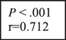
In addition, a good degree of correlation was determined between the elevation values in patients’ cerebral and abdominal saturations. A good degree of correlation was also present between elevation values in the right cerebral saturation and right abdominal saturation (r = 0.712). A good degree of correlation was also present between the left cerebral saturation and left abdominal saturation (r = 0.699) (Figs. 1 and 2).
- Discussion
Patients with prehospitalization CA and elevated abdominal tissue saturation had a higher level of ROSC compared to those with low saturation. Circulation did not return in patients with low abdominal tis- sue saturations. We also determined a good correlation between abdom- inal saturations and cerebral saturations of patients with out-of-hospital CA with or without spontaneous circulation return. Based on the results of our study, we can assume that the measurement of abdominal satura- tion can be indicative of the ROSC. This is the first study to assess abdom- inal saturation during CPR using a noninvasive technique.
Although there are several studies assessing abdominal saturation with NIRS in various diseases, there are no studies evaluating this during CPR [9,11,16-19]. One animal model study performed to determine
Receiver operating characteristic analyses of patient parameters
|
Parameters |
Cutoff |
Alive |
Deceased |
Odds ratio (95% CI) |
AUC |
Performance |
|
Age |
<=66 |
11 |
9 |
7.33 (1.291-41.652) |
0.714 |
Sens: 57.1% |
|
N 66 |
2 |
12 |
P = .038 |
Spec: 84.6% |
||
|
Length of CPR (min) |
<=22 |
13 |
1 |
14.08 (2.119-90.909) |
0.996 |
Sens: 95.2% |
|
N 22 |
0 |
20 |
P b .001 |
Spec: 100% |
||
|
Mean right ScO2 on dischargea |
N 35 |
13 |
1 |
14.08 (2.119-90.909) |
0.998 |
Sens: 95.2% |
|
<=35 |
0 |
20 |
P b .001 |
Spec: 100% |
||
|
Mean right ?ScO2 |
N 18 |
12 |
0 |
22 (3.242-149.298) |
0.995 |
Sens: 100% |
|
<=18 |
1 |
21 |
P b .001 |
Spec: 92.3% |
||
|
Mean left ScO2 on discharge |
N 37 |
13 |
1 |
14.08 (2.119-90.909) |
0.998 |
Sens: 95.2% |
|
<=37 |
0 |
20 |
P b .001 |
Spec: 100% |
||
|
Mean left ?ScO2 |
N 20 |
12 |
0 |
22 (3.242-149.298) |
0.984 |
Sens: 100% |
|
<=20 |
1 |
21 |
P b .001 |
Spec: 92.3% |
||
|
Mean right rSO2 on discharge |
N 40 |
13 |
0 |
– |
1.00 |
Sens: 100% |
|
<=40 |
0 |
21 |
P b .001 |
Spec: 100% |
||
|
Mean right ?rSO2 |
N 4 |
13 |
0 |
– |
1.00 |
Sens: 100% |
|
<=4 |
0 |
21 |
P b .001 |
Spec: 100% |
||
|
Mean left rSO2 on discharge |
N 51 |
13 |
0 |
– |
1.00 |
Sens: 100% |
|
<=51 |
0 |
21 |
P b .001 |
Spec: 100% |
||
|
Mean left ?rSO2 |
N 5 |
13 |
0 |
– |
1.00 |
Sens: 100% |
|
<=5 |
0 |
21 |
P b .001 |
Spec: 100% |
Abbreviations: CI, confidence interval; Sens, sensitivity; Spec, specificity.
a Discharge means admission of the patient to intensive care unit after ROSC.
60
Increase of right rSO2
50
40
30
20
10
0
-10


0
10
20
30
40
50
60
70
80
90
Increase of right ScO2
Fig. 1. Correlation between right ?rSO2 and right ?ScO2.
abdominal ischemia in patients with shock compared invasive techniques with NIRS and reported that NIRS was a good means of detecting abdominal ischemia. In that study, hemorrhagic shock was experimentally induced in piglets, and hepatic and intestinal blood flow was determined to decrease. Near-infrared spectrophotometry was reported to be a particularly good method for predicting abdominal ischemia [20]. Abdominal O2 saturation values at the beginning of CPR were low in almost all our patients admitted with CA, which were regarded as abdominal ischemia. Spontaneous circulation returned in patients whose rSO2 measurements subsequently began to rise.
Most studies, in which cerebral oxygenation was monitored by NIRS during CPR, show that an increase in the low values measured at the onset of resuscitation is positively correlated with the ROSC [4,21]. Ahn et al [7] measured cerebral oxygenation in their patients with out-of-hospital CA during CPR. Return of spontaneous circula- tion was observed in patients with increasing ScO2 levels over the initial values [7].
In our study, we observed that spontaneous circulation returned in patients with elevated abdominal saturation. Determination of both ce- rebral and abdominal saturations simultaneously renders our study su- perior to that of Ahn et al [7]. Because there was a good correlation between cerebral and abdominal oxygenations, elevation of abdominal saturation is also a good indicator of ROSC.
Frisch et al [22] measured prehospital tissue saturations of patients with CA using NIRS in a series of 5 patients and suggested that patients with low values were at greater risk for rearrest. They compared tissue saturation and end-tidal CO2 and reported that NIRS measure- ments predict arrest in an earlier period. Because tissue saturation mea- surement can predict rearrest, abdominal saturation establishment can predict ROSC.
In a previous study, we determined that spontaneous circulation returned in patients with elevating cerebral saturation during CPR. In this study, we evaluated both the cerebral and abdominal saturations of the patients. We found a good degree of correlation between cerebral and abdominal saturations, which supports our previous study. The im- plication in both studies is that abdominal saturation determination alone can be indicative of ROSC [23].
Ischemic status in conditions in which abdominal pressure in- creases, known as Abdominal compartment syndrome, can be deter- mined using NIRS. Varela et al [19] suggested that NIRS can show early changes in mesenteric and systemic perfusion. They measured ab- dominal circulation using invasive methods and NIRS. They determined no significant difference between the invasive methods and NIRS in measuring abdominal saturation and thus recommended the use of NIRS [19]. This study supports the use of NIRS in the measurement of ab- dominal saturation. It also supports our hypothesis in that changes in the mesenteric perfusions affect the systemic perfusion, whose varia- tions at the earlier stages can be picked up by NIRS. Monitoring of ab- dominal saturation during CPR is indicative of return or otherwise of spontaneous circulation.
The findings of the study of Li et al [24] were also in line with our own. They evaluated cerebral and abdominal saturations in patients using NIRS with the Norwood procedure (a surgical technique applied in patients with congenital heart diseases). They determined that both cerebral and abdominal perfusions decreased in conditions in which systemic perfusion was compromised. They also supported the idea of using NIRS, a noninvasive measurement technique predicting the im- provement of abdominal perfusion [24]. This is not surprising because NIRS measures oxyhemoglobin and deoxyhemoglobin in circulation, and measurement values will fall if circulation is compromised. Initial
70


0
10
20
30
40
50
60
70
80
60
50
Increase of left rSO2
40
30
20
10
0
-10
Increase of left ScO2
Fig. 2. Correlation between left ?rSO2 and left ?ScO2.
measurements in patients with arrest were also low in our study, rising when spontaneous circulation was restored and systemic blood pres- sure was established.
Studies focusing on the circulation of Major organs during CPR are on the increase [25-29]. In this regard, although monitoring of cerebral saturation by NIRS has been projected as the only aid in determining ROSC, it has become apparent that monitoring of abdominal saturation can serve the same purpose.
In light of all these studies, we think that the continuous monitoring of patients’ abdominal saturation during CPR is useful in predicting both ROSC and Ischemic injury in the splanchnic region.
- Limitations
The low patient number is the main limitation of this study. Future multicenter studies with a larger patient population are required. In addition, long-term monitoring of abdominal saturation of the patients whose spontaneous circulation has been established is lacking. Although abdominal saturation monitoring is correlated with the ROSC, its effect on hospital discharge is unknown. A biochemical marker study may be warranted on those patients whose spontaneous circula- tion has returned to detect possible ischaemic injuries in the long term.
- Conclusions
The best way of increasing postarrest discharge is still basic life sup- port, and optimal application of advanced cardiac life support rules. Pa- tients with increased abdominal and cerebral saturation values have a higher survival rate after appropriate CPR. This noninvasive measure- ment system and monitoring of patients during CPR may be a good method of predicting ROSC and assessing abdominal perfusion.
- Newman DH, Callaway CW, Greenwald IB, Freed J. Cerebral oximetry in out-of hospital cardiac arrest: standard CPR rarely provides detectable hemoglobin- oxygen saturation to the frontal cortex. Resuscitation 2004;63:189-94.
- Parnia S, Nasir A, Shah C, Patel R, Mani A, Richman P. A feasibility study evaluating the role of cerebral oximetry in predicting return of spontaneous circulation in cardiac arrest. Resuscitation 2012;83:982-5.
- Kamarainen A, Sainio M, Olkkola KT, Huhtala H, Tenhunen J, Hoppu S. Quality controlled manual chest compressions and cerebral oxygenation during in- hospital cardiac arrest. Resuscitation 2012;83:138-42.
- Meex I, De Deyne C, Dens J, Scheyltjens S, Lathouwers K, Boer W, et al. Feasibility of absolute cerebral tissue oxygen saturation during cardiopulmonary resuscitation. Crit Care 2013;17(2):R36.
- Singer AJ, Ahn A, Inigo-Santiago LA, Thode Jr HC, Henry MC, Parnia S. Cerebral oxim- etry levels during CPR are associated with return of spontaneous circulation follow- ing cardiac arrest: an observational study. Emerg Med J 2014 Mar 24. http://dx.doi. org/10.1136/emermed-2013-203467 [Epub ahead of print].
- Koyama Y, Wada T, Lohman BD, Takamatsu Y, Matsumoto J, Fujitani S, et al. A new method to detect cerebral blood flow waveform in synchrony with chest compression by Near-infrared spectroscopy during CPR. Am J Emerg Med 2013;31:1504-8.
- Ahn A, Nasir A, Malik H, D’Orazi F, Parnia S. A pilot study examining the role of regional cerebral oxygen saturation monitoring as a marker of return of spontane-
ous circulation in shockable (VF/VT) and non-shockable (PEA/Asystole) causes of cardiac arrest. Resuscitation 2013;84:1713-6.
- Ito N, Nanto S, Nagao K, Hatanaka T, Kai T. Regional cerebral oxygen saturation predicts poor neurological outcome in patients with out-of-hospital cardiac arrest. Resuscitation 2010;81:1736-7.
- Westgarth-Taylor C, de Lijster L, van Bogerijen G, Millar AJ, Karpelowsky J. A prospective assessment of renal oxygenation in children undergoing laparoscopy using near-infrared spectroscopy. Surg Endosc 2013;27:3696-704.
- Gillam-Krakauer M, Cochran CM, Slaughter JC, Polavarapu S, McElroy SJ, Hernanz- Schulman M, et al. Correlation of abdominal rSO2 with superior mesenteric artery velocities in preterm infants. J Perinatol 2013;33:609-12.
- Kaufman J, Almodovar MC, Zuk J, Friesen RH. Correlation of abdominal site near- infrared spectroscopy with gastric tonometry in infants following surgery for con- genital heart disease. Pediatr Crit Care Med 2008;9:62-8.
- Nollert G, Jonas R, Reichart B. Optimizing cerebral oxygenation during cardiac sur- gery: a review of experimental and clinical investigations with near infrared spec- trophotometry. Thorac Cardiovasc Surg 2000;48:247-53.
- Denault A, Deschamps A, Murkin J. Cerebral oximetry monitoring in anesthesiology. Anesthesiol Rounds 2008;7(2).
- Davie SN, Grocott HP. Impact of extracranial contamination on regional cerebral oxygen saturation. Anesthesiology 2012;116:834-40.
- Edmonds Jr HL, Ganzel BL, Austin III EH. Cerebral oximetry for cardiac and vascular surgery. Semin Cardiothorac Vasc Anesth 2004;8:147-66.
- Nahum E, Skippen PW, Gagnon RE, Macnab AJ, Skarsgard ED. Correlation of near- infrared spectroscopy with Perfusion parameters at the hepatic and systemic levels in an endotoxemic shock model. Med Sci Monit 2006;12:313-7.
- Said MM, Niforatos N, Rais-Bahrami K. Validation of near infrared spectroscopy to measure abdominal somatic tissue oxygen saturation in neonates. J Neonatal Perinat Med 2013;6:23-30.
- Demirel G, Oguz SS, Celik IH, Erdeve O, Dilmen U. Cerebral and mesenteric tissue oxygenation by positional changes in very low birth weight premature infants. Early Hum Dev 2012;88:409-11.
- Varela JE, Cohn SM, Giannotti GD, Dolich MO, Ramon H, Wiseberg JA, et al. Near- infrared spectroscopy reflects changes in mesenteric and systemic perfusion during abdominal compartment syndrome. Surgery 2001;129:363-70.
- Cohn SM, Varela JE, Giannotti G, Dolich MO, Brown M, Feinstein A, et al. Splanchnic perfusion evaluation during hemorrhage and resuscitation with gastric near- infrared spectroscopy. J Trauma 2001;50:629-34.
- Reynolds JC, Salcido D, Koller AC, Sundermann ML, Frisch A, Suffoletto BP, et al. Tissue oximetry by near-infrared spectroscopy in a porcine model of out-of- hospital cardiac arrest and resuscitation. Resuscitation 2013;84:843-7.
- Frisch A, Suffoletto BP, Frank R, Martin-Gill C, Menegazzi JJ. Potential utility of near- infrared spectroscopy in out-of-hospital cardiac arrest: an illustrative case series. Prehosp Emerg Care 2012;16:564-70.
- Asim K, Gokhan E, Ozlem B, et al. Near infrared spectrophotometry (cerebral oxim- etry) in predicting the return of spontaneous circulation in out-of-hospital cardiac arrest. Am J Emerg Med 2014;32:14-7.
- Li J, Van Arsdell GS, Zhang G, Cai S, Humpl T, Caldarone CA, et al. Assessment of the relationship between cerebral and splanchnic oxygen saturations measured by near- infrared spectroscopy and direct measurements of systemic haemodynamic vari- ables and oxygen transport after the Norwood procedure. Heart 2006;92:1678-85.
- Storm C, Leithner C, Krannich A, Wutzler A, Ploner CJ, Trenkmann L, et al. Regional cerebral oxygen saturation after cardiac arrest in 60 patients a prospective outcome study. Resuscitation 2014;85:1037-41.
- Genbrugge C, Dens J, Meex I, Boer W, Jans F, De Deyne C. Cerebral saturation moni- toring during cardiopulmonary resuscitation should be used as dynamic, rather than static, information. Resuscitation 2013;84:e111-2.
- Loukas T, Vasileiadis I, Anastasiou H, Karatzanos E, Gerovasili V, Nana E, et al. Resuscitation after cardiac arrest in a septic porcine model: adding vasopressin vs epinephrine alone administration. BMC Res Notes 2014;7:49.
- Parnia S, Nasir A, Ahn A, Malik H, Yang J, Zhu J, et al. A feasibility study of cerebral oximetry during in-hospital mechanical and Manual cardiopulmonary resuscitation. Crit Care Med 2014;42:930-3.
- Schewe JC, Thudium MO, Kappler J, Steinhagen F, Eichhorn L, Erdfelder F, et al. Mon- itoring of cerebral oxygen saturation during resuscitation in out-of-hospital cardiac arrest: a feasibility study in a physician staffed emergency medical system. Scand J Trauma Resusc Emerg Med 2014;22:58.